Knee Ligament Injuries
These articles are for general information only and are not medical advice. Full Disclaimer. All articles compliments of the AAOS
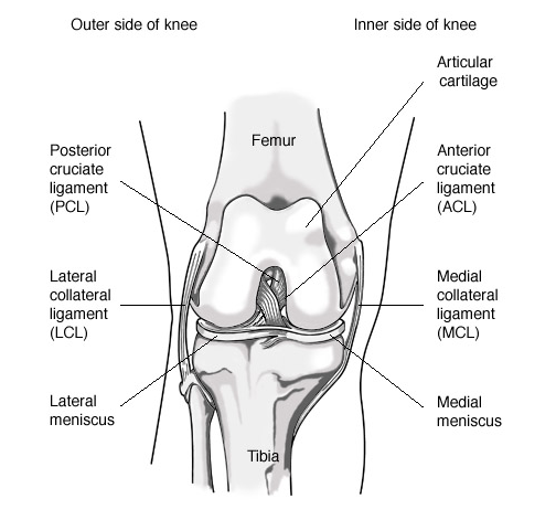
In 2003 more than 9.5 million people visited orthopedic surgeons because of knee problems. (Source: National Center for Health Statistics; Centers for Disease Control and Prevention; 2003 National Ambulatory Medical Care Survey.) The knee is the largest joint in the body and is vital to movement. Two sets of ligaments in the knee give it stability: the cruciate and the collateral ligaments.
Cruciate Ligaments
The cruciate ligaments are located inside the knee joint and connect the thighbone (femur) to the shinbone (tibia). They are made of many strands and function like short ropes that hold the knee joint tightly in place when the leg is bent or straight. This stability is needed for proper knee joint movement.
The name, cruciate, derives from the word crux, meaning cross, and crucial. The cruciate ligaments not only lie inside the knee joint, they crisscross each other to form an “x”. The cruciate ligament located toward the front of the knee is the anterior cruciate ligament (ACL), and the one located toward the rear of the knee is called the posterior cruciate ligament (PCL).
ACL Injuries
The ACL prevents the shinbone from sliding forwards beneath the thighbone. The ACL can be injured in several ways:
- Changing direction rapidly
- Slowing down when running
- Landing from a jump
- Direct contact, such as in a football tackle
Recognizing an ACL Injury
If you injure your ACL, you may not feel any pain immediately. However, you might hear a popping noise and feel your knee give out from under you. Within 2 to 12 hours, the knee will swell, and you will feel pain when you try to stand. Apply ice to control swelling and elevate your knee until you can see an orthopedic surgeon.
If you walk or run on an injured ACL, you can damage the cushioning cartilage in the knee. For example, you may plant the foot and turn the body to pivot, only to have the shinbone stay in place as the thighbone above it moves with the body.
Diagnosing an ACL Injury
A diagnosis of ACL injury is based on a thorough physical examination of the knee. The exam may include several tests to see if the knee stays in the proper position when pressure is applied from different directions. Your orthopaedist may order an X-ray and MRI (magnetic resonance imaging) or, in some cases, arthroscopic inspection.
A partial tear of the ACL may or may not require surgical treatment. A complete tear is more serious. Complete tears, especially in younger athletes, may require reconstruction.
Treating ACL Tears
Both nonoperative and operative treatment choices are available.
- Nonoperative treatment:
- May be used because of a patient’s age or overall low activity level
- May be recommended if the overall stability of the knee seems good
- Involves a treatment program of muscle strengthening, often with the use of a brace to provide stability
- Operative treatment (either arthroscopic or open surgery): Uses a strip of tendon, usually taken from the patient’s knee (patellar tendon) or hamstring muscle, that is passed through the inside of the joint and secured to the thighbone and shinbone
- Is followed by an exercise and rehabilitation program to strengthen the muscles and restore full joint mobility
PCL Injuries
The posterior cruciate ligament, or PCL, is not injured as frequently as the ACL. PCL sprains usually occur because the ligament was pulled or stretched too far, a blow to the front of the knee, or a simple misstep.
PCL injuries disrupt knee joint stability because the shinbone can sag backwards. The ends of the thighbone and shinbone rub directly against each other, causing wear and tear to the thin, smooth articular cartilage. This abrasion may lead to arthritis in the knee.
Treating PCL Injuries
Patients with PCL tears often do not have symptoms of instability in their knees, so surgery is not always needed. Many athletes return to activity without significant impairment after completing a prescribed rehabilitation program.
However, if the PCL injury pulls a piece of bone out of the top of the shinbone, surgery is needed to reattach the ligament. Knee function after this surgery is often quite good.
Collateral Ligaments
The collateral ligaments are located at the inner side and outer side of the knee joint. The medial collateral ligament (MCL) connects the thighbone to the shinbone and provides stability to the inner side of the knee. The lateral collateral ligament (LCL) connects the thighbone to the other bone in the lower portion of your leg (fibula) and stabilizes the outer side.
Injuries to the MCL are usually caused by contact on the outside of the knee and are accompanied by sharp pain on the inside of the knee. The LCL is rarely injured.
Collateral Ligament Injuries
If the medial collateral ligament (MCL) has a small partial tear, conservative treatment usually works. Remember the acronym RICE: Rest, Ice, Compression, Elevation.
Rest the knee to give the ligament time to heal. Ice can be applied two or three times a day for 15 to 20 minutes each time.
Compress the injury to limit swelling. You may have to wear a bandage or brace for a while.
Elevate the knee whenever possible.
You should also consult your physician about a course of rehabilitation exercises for good healing.
If the collateral ligament is completely torn or torn in such a way that ligament fibers cannot heal, you may need surgery. Repair may bring good results, with a return to good knee stability. After satisfactory rehabilitation, many people resume their previous levels of activity.
A rehabilitation plan is needed if you have a cruciate or collateral ligament injury. Most rehabilitation plans include:
- Passive range-of-motion exercises designed to restore flexibility
- Braces to control joint movement
- Exercises to strengthen the quadriceps muscles in the front of the thigh. (Muscle strength is needed to provide the knee joint with as much support and stability as possible when weight is placed on it.)
- Additional exercises on a high-seat exercise bicycle, followed by more strenuous quadriceps exercise
Your progress and the ability of the knee to function as a normal knee will determine how long you must use crutches and a brace.

