Developmental Dislocation (Dysplasia) of the Hip (DDH)
Developmental dislocation of the hip (DDH) is an abnormal formation of the hip joint. The ball at the top of the thighbone (femoral head) is not stable in the socket (acetabulum). Also, the ligaments of the hip joint may be loose and stretched. The degree of instability or looseness varies. A baby born with DDH may have the ball of his or her hip loosely in the socket, the looseness may worsen as the child grows and becomes more active, or the ball may be completely dislocated at birth. It is important to note the ball may not always be out of the socket at birth.
Hip dysplasia is often noted in the newborn exam. Treatment is easier and safer the earlier the diagnosis is made. Hips found normal at birth can be found abnormal later, but this is rare. Pediatricians screen for DDH at a newborn’s first exam and at every well-baby checkup thereafter. A child’s hip may not be dislocated at birth, which means the condition may not be noticed until a child begins to walk, by which time treatment is more complicated and uncertain.

Left untreated, DDH or hip dysplasia leads to pain and osteoarthritis by early adulthood. It may cause legs of different lengths or a “duck-like” walk and decreased agility. If dysplasia is treated successfully (and the earlier the better) children end up with normal hip joint function, have no further problems and go on to lead active lives. However, even with appropriate treatment, especially in the child who is 2 years or older, hip deformity and osteoarthritis may develop later in life.
Risk Factors / Prevention
DDH has a familial tendency. It can be present in either hip and in any individual. It usually affects the left hip and is predominant in:
- First born children.
- Babies born in the breech position (especially with feet up by the shoulders). The American Academy of Pediatrics now recommends ultrasound screening of all female, breech babies after birth to show if DDH is present.
Symptoms
Although some dislocated hips show no signs, contact a doctor if your baby has
- Legs of different lengths.
- Uneven thigh skin folds.
- Less mobility or flexibility on one side.
In children who have begun to walk, limping, toe walking and a waddling “duck-like” gait are also signs.
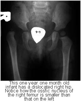
In addition to visual clues, doctors use careful physical examination tests to check for subtle signs of hip instability or dislocation in babies, such as listening and feeling for “clunks.” Hip X-rays also may be helpful in older infants and children.

Treatment Options
Treatment methods depend upon the child’s age.
Newborn
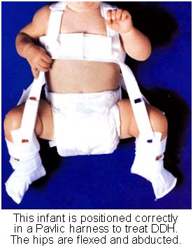
An unstable hip recognized at birth is treated with a soft, simple positioning device (Pavlik harness) for one or two months to keep the hip bone in its socket. This may help tighten ligaments and stimulate normal hip socket formation.
1-6 months
Treatment to reposition the hip ball in the socket uses a harness or similar device. The method is usually successful; if it is not, the joint may be positioned into place under anesthesia (closed reduction) and maintained with a body cast (spica).
6 months-2 years
Manipulation of the socket under anesthesia (closed reduction) is the major method of treatment. Open surgery may be necessary. Both require a body cast (spica).
After 2 years
Deformities may have become severe, making major open surgical intervention necessary to realign the hip. This is followed by a body cast (spica).
The child will need a body cast and/or brace to keep his or her hip bone in the joint while healing after operations. X-rays and other regular follow-up monitoring are needed after DDH treatment until the child’s growth is complete. Complications may include a small delay in the development of walking if he or she uses a cast. Positioning devices may cause skin irritation, and a difference in leg lengths may remain. Growth disturbance of the upper thigh rarely occurs.
